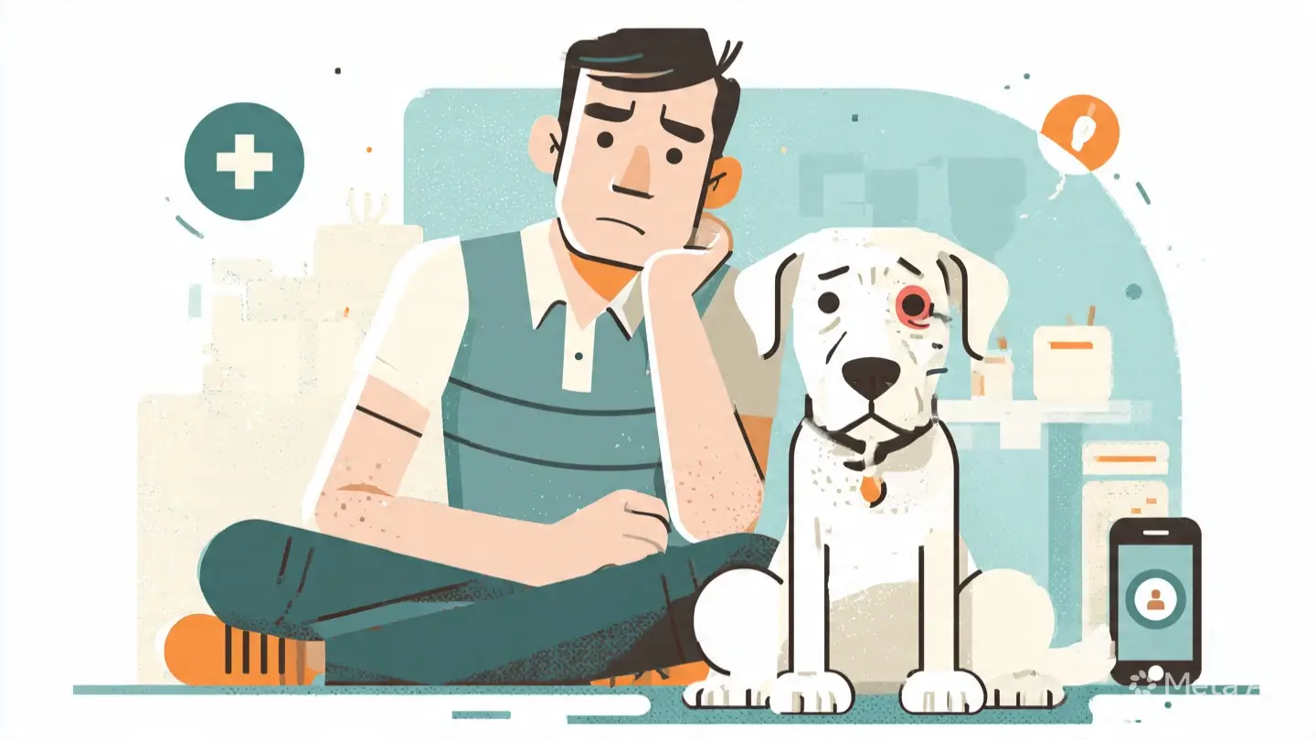
Cherry eye is one of the more visually striking conditions a dog owner can encounter — a smooth, round, reddish-pink mass appearing suddenly in the inner corner of the eye, looking somewhat like a cherry. While alarming in appearance, cherry eye is generally not a painful emergency, but it does require veterinary treatment to prevent long-term complications.
If your dog has developed a visible mass in the corner of their eye, contact Walker Lake Veterinary Centre at (587) 786-6771 for an assessment.
Cherry eye is not a tumour or growth. It is the prolapse — or displacement — of the tear-producing gland of the third eyelid from its normal position. Understanding what has actually happened helps make sense of both the appearance and the treatment.
Understanding the Third Eyelid and Its Gland
Dogs — unlike humans — have three eyelids. The third eyelid, also called the nictitating membrane, is a translucent membrane located in the inner corner of the eye. It sweeps across the eye surface to distribute the tear film, remove debris, and provide additional protection.
Embedded within this third eyelid is the nictitans gland — a tear-producing gland that contributes approximately 30 to 50% of the total tear production in a dog’s eye. This gland is normally anchored firmly within the third eyelid by connective tissue.
In cherry eye, the connective tissue anchor fails and the gland prolapse — it flips out of its normal position and becomes visible as the characteristic pink-red mass at the inner corner of the eye.
What Causes Cherry Eye?
The exact cause of the connective tissue weakness that allows the gland to prolapse is not fully understood. It is believed to have a significant hereditary component — certain breeds have a known predisposition that suggests a genetic weakness in the connective tissue supporting the gland.
Predisposed Breeds
Cherry eye is significantly more common in certain breeds:
- Bulldogs and French Bulldogs — among the most commonly affected
- Cocker Spaniels
- Beagles
- Basset Hounds
- Shih Tzus
- Lhasa Apsos
- Poodles
- Great Danes
- Neapolitan Mastiffs
- Boston Terriers
Age of Onset
Cherry eye typically occurs in young dogs — most commonly under two years of age. This is consistent with the idea that the connective tissue anchor is inherently weak in predisposed individuals and fails as the dog grows.
Bilateral Involvement
Cherry eye frequently affects both eyes — either simultaneously or with the second eye developing the condition weeks to months after the first. Dogs that develop cherry eye in one eye have a significant likelihood of developing it in the other. This has implications for treatment planning and owner expectations.
Signs and Symptoms of Cherry Eye
The Prolapsed Gland
The most obvious sign is the appearance of a smooth, round, pink to red mass in the medial corner — the inner corner — of one or both eyes. The mass may be:
- Constantly visible
- Intermittently visible — disappearing and reappearing as the gland slips in and out of position
- Small and barely noticeable, or large enough to cover a significant portion of the eye
Secondary Signs
Once the gland is prolapsed, it is exposed to air and environmental irritants rather than being protected within the third eyelid. This causes:
- Redness and swelling of the prolapsed gland — it becomes increasingly inflamed the longer it remains exposed
- Ocular discharge — mucoid or mucopurulent discharge from the eye
- Excessive tearing
- Squinting or pawing at the eye — indicating discomfort
- Conjunctivitis — inflammation of the surrounding eye tissue
- Dry eye development over time if the gland is damaged or removed
⚠️ Do not attempt to manually push the prolapsed gland back into position at home. This rarely succeeds, risks damaging the gland, and delays appropriate veterinary treatment.
Why Prompt Treatment Matters
The prolapsed gland is a functioning tear-producing structure that contributes significantly to the eye’s total tear production. The longer it remains exposed and inflamed, the greater the risk of permanent damage to the gland tissue.
Damaged gland tissue produces less tears. A dog that loses significant tear-producing capacity — particularly from both eyes — is at risk of developing keratoconjunctivitis sicca (KCS), commonly known as dry eye. Dry eye is a chronic, uncomfortable condition that requires lifelong management and can eventually lead to corneal scarring and vision impairment.
This is why early surgical treatment — before the gland becomes significantly inflamed and damaged — gives the best long-term outcome.
Treatment Options for Cherry Eye
Surgical Repositioning — The Recommended Approach
The gold standard treatment for cherry eye is surgical repositioning of the prolapsed gland back into its correct anatomical position within the third eyelid. This preserves the gland’s tear-producing function — critically important for long-term eye health.
Several surgical techniques exist for repositioning the gland. The two most commonly used are:
The Pocket Technique
A small pocket is created within the third eyelid tissue and the prolapsed gland is tucked inside and secured with sutures. The pocket technique is widely used and has a good success rate. It preserves the gland completely.
The Imbrication Technique
Sutures are placed to tighten the connective tissue anchoring the gland, pulling it back into position. This technique is also effective and preserves the gland.
Success rates for surgical repositioning vary between studies but are generally reported at 80 to 95%. Some dogs — particularly brachycephalic breeds with flat faces — have higher recurrence rates and may require revision surgery.
Surgical Removal — Not Recommended
Historically, the standard treatment for cherry eye was simply to remove the prolapsed gland. This is now strongly discouraged by veterinary ophthalmologists because removing the gland eliminates 30 to 50% of tear production, dramatically increasing the risk of dry eye — particularly when the second eye subsequently develops cherry eye.
Surgical removal may still be considered in a small number of cases where the gland is too severely damaged to be repositioned effectively, but this should only be done after careful discussion of the long-term implications.
If your dog has had cherry eye treated by gland removal in the past — perhaps before the risks were fully understood or before repositioning techniques became standard — discuss dry eye monitoring with your veterinarian. Regular tear production testing is important for these dogs.
Temporary Medical Management
While waiting for surgery, topical anti-inflammatory eye drops may be prescribed to reduce swelling of the prolapsed gland and make subsequent surgical repositioning more straightforward. Medical management alone does not resolve cherry eye — the gland will not return to its correct position without surgical intervention.
What to Expect After Cherry Eye Surgery
Immediate Post-Operative Period
After surgery your dog will typically go home the same day with:
- An Elizabethan collar (e-collar) — essential to prevent pawing at the eye
- Topical antibiotic and anti-inflammatory eye drops — applied several times daily
- Oral anti-inflammatory medication in some cases
- Clear written aftercare instructions
Recovery Timeline
Most dogs recover well within 2 to 3 weeks of surgery. During this period:
- The e-collar must remain on at all times — even for a few minutes of unsupervised access can result in self-trauma that damages the surgical repair
- Eye drops must be applied as prescribed — consistency is essential for reducing inflammation and preventing infection
- Avoid bathing, swimming, or exposing the eye to water
- Keep your dog calm and restrict vigorous exercise and rough play
- Attend all scheduled follow-up appointments
Recurrence
Despite successful surgery, cherry eye can recur — the gland prolapses again. Recurrence rates vary between breeds and surgical techniques. Brachycephalic breeds have higher recurrence rates. If recurrence occurs, revision surgery using the same or an alternative technique is typically performed.
The Second Eye
As discussed above, many dogs that develop cherry eye in one eye will develop it in the other eye subsequently. This is not a failure of the surgical repair — it reflects the bilateral nature of the underlying connective tissue weakness. Owners of predisposed breeds should be aware of this possibility and monitor the second eye closely.
Long-Term Eye Health After Cherry Eye
Dry Eye Monitoring
Even dogs that have successful gland repositioning benefit from periodic tear production testing — the Schirmer Tear Test measures tear production volume and takes only a few minutes to perform. This is particularly important if the gland was noted to be significantly inflamed at the time of surgery, or if the dog develops cherry eye in the second eye.
Signs of Dry Eye to Watch For
- Thick, mucoid or mucopurulent eye discharge
- Dull, hazy appearance to the corneal surface
- Redness and chronic eye irritation
- Squinting and pawing at the eye
- Progressive corneal pigmentation — dark discolouration of the eye surface
If any of these signs develop, a veterinary eye examination and tear production test should be scheduled promptly.
If your dog has developed cherry eye or any eye concern, book an appointment at Walker Lake Veterinary Centre. Call (587) 786-6771 or book online. Located at 5109 22 Avenue SW, Southwest Edmonton.
Book an Appointment at Walker Lake Veterinary Centre
Walker Lake Veterinary Centre provides eye examination, cherry eye assessment, and surgical treatment for dogs and cats across Walker Lake, Summerside, Ellerslie, Heritage Valley, and Southwest Edmonton.
Call us at (587) 786-6771 or book online. We are located at 5109 22 Avenue SW, Edmonton.
Clinic hours:
- Monday, Tuesday & Thursday: 9:00 AM – 7:00 PM
- Wednesday & Friday: 9:00 AM – 6:00 PM
- Saturday: 9:00 AM – 3:00 PM
- Sunday: Closed
Frequently Asked Questions
Is cherry eye painful for dogs?
Cherry eye is not typically acutely painful in the way that a wound or injury is painful. However the exposed, inflamed gland causes significant ocular discomfort — most dogs with cherry eye rub and paw at the affected eye. The associated conjunctivitis and dry eye that can develop if left untreated cause ongoing discomfort. Early treatment prevents unnecessary suffering.
Can cherry eye resolve on its own?
Very rarely and unpredictably. Occasional spontaneous resolution has been reported in very mild cases — particularly when the gland intermittently prolapses and returns to position on its own. However, waiting for spontaneous resolution risks progressive inflammation and damage to the gland. Surgical treatment is recommended for all persistent cherry eye.
How much does cherry eye surgery cost?
The cost of cherry eye surgery depends on whether one or both eyes are involved, the surgical technique used, and whether general anaesthesia or heavy sedation is required. Contact Walker Lake Veterinary Centre at (587) 786-6771 for a specific quote based on your dog’s individual situation.
My dog had cherry eye surgery but it has come back — what now?
Recurrence after surgery is a known possibility, particularly in brachycephalic breeds. Revision surgery using the same or an alternative repositioning technique is typically performed. Discuss the options with your veterinarian — the goal remains preserving the gland’s tear-producing function wherever possible.
Can I prevent cherry eye in my dog?
Because cherry eye has a strong hereditary component, it cannot be reliably prevented in predisposed breeds. Responsible breeding — selecting breeding dogs without a history of cherry eye — can reduce prevalence over generations. For individual owners of predisposed breeds, early veterinary assessment of any eye abnormality and prompt treatment when cherry eye occurs are the most important steps.